MCH Radiology Department
Nuclear Medicine
As of 2023, MCH is proud to offer in house nuclear medicine!
General Nuclear Medicine:
Nuclear medicine comprises diagnostic examinations that result in images of body anatomy and function. The images are developed based on the detection of energy emitted from a radioactive substance given to the patient, either intravenously or by mouth. Generally, radiation to the patient is similar to that resulting from standard x-ray examinations.
Cardiac Nuclear Medicine:
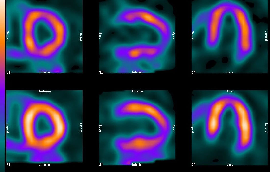
A nuclear stress test is a procedure that checks the blood flow to the heart and how well your heart is beating. You will receive a small, safe amount of radioactive medication that circulates to the heart muscle. A special camera is used to measure how well this medication spreads throughout the heart.
Hours
The following exams are available every Thursday:
-
Bone Imaging
-
Cardiac Blood Pool Single
-
Cardiac Rest/Stress Testing
-
Gastric Emptying Study
-
Gastrointestinal Blood Loss Imaging
-
Hepatobiliary Imaging
-
Intestine Imaging
-
Kidney Imaging
-
Parathyroid Study
-
Pulmonary Perfusion Imaging
-
Thyroid Imaging
An appointment is required for these exams and start time is dependent on the exam being performed.
Frequently Asked Questions...
How Do I Prepare for a Nuclear Study?
Some Nuclear Studies have a prep and some do not. When scheduling your appointment, you will be informed of any prep, we will also call you to remind you of your prep the day prior to your exam.
What Does the Equipment Look Like?
During most nuclear medicine examinations, you will lie down on a scanning table. Consequently, the only piece of equipment you may notice is the specialized nuclear imaging camera used during the procedure. It is enclosed in metallic housing designed to facilitate imaging of specific parts of the body. It can look like a large round metallic apparatus suspended from a tall, moveable post or a sleek one-piece metal arm that hangs over the examination table. A nearby computer console processes the data from the procedure.
How Does the Procedure Work?
You are given a small dose of radioactive material, usually intravenously but sometimes orally, that localizes in specific body organ systems. This compound, called a radiopharmaceutical agent or tracer, eventually collects in the organ and gives off energy as gamma rays. The gamma camera detects the rays and works with a computer to produce images and measurements of organs and tissues.
We offer online sharing of your medical images using Nuance Powershare. This service allows us to share images with your provider in a secure and convenient way!
All imaging will be sent to the physicians of Manhattan Radiology to be read and reported. Your ordering provider will have your report within 24 to 48 hours. Your provider should contact you with your results.
All services require a written order from a physician in good standing and licensed to practice medicine in the State of Kansas.

